The main activities in this field are related to the digital radiography and tomography also of large objects. The research started in 2008, in collaboration with the Physics Department
of the University of Bologna and the Conservation and Restoration Center "La Venaria Reale". The main aim of this research is the development of imaging setups able to analyse
artworks and archaeological finds. Since in this field the dimensions and materials are heterogeneous, one instrument cannot suite all the cases.For this reason up to now two systems were developed, both able to perform radiographs and tomographs:
the first, installed at the CCR "La Venaria Reale" and developed in the framework of the neu_ART regional project, allows for the 2D analysis of large paintings (up to 3.1 m x 2.7 m)
and 3D of large statues or furniture (with height of up to 2.5 m and width of up to 2 m); the second, in the laboratories of the Physics Department, allow for the analysis of smaller objects,
but with a better resolution. Recently a new apparatus was developed in the framework of the SAX-Infra-P (POR FESR) project; the particularlty of the apparatus
is the use of a liquid anode X-ray source, able to achieve significantly higher brightness and smaller spot sizes than any other available microfocus X-ray source.
These techniques are based on the different x-rays absorption of an object, due both to different thickness of the same material or to the presence of different materials.
The obtainable results are grey scale images, in 2D for radiography and in 3D for tomography, that can give useful information about:
- differences among constituent materials - alterations and state of preservation - hidden features - constructive techniques and previous restoration
All this information increases the knowledge about an artwork or an archaeological find and can be useful for art-historians, archaeologists and restorers,
allowing to see inside an object in a totally non-invasive way. Analysis were also carried out by means of synchrotron radiation at Elettra in Trieste.
Other than conventional digital 2D radiography and tomography, an apparatus for differential radiography able to perform quantitative two-dimensional mapping of single elements on paintings was developed.
The apparatus has been designed on the basis of the expertise coming from previous research projects funded by Ferrara University and INFN division of Ferrara and it has been built
and finalized at the INFN technological workshop in Torino.
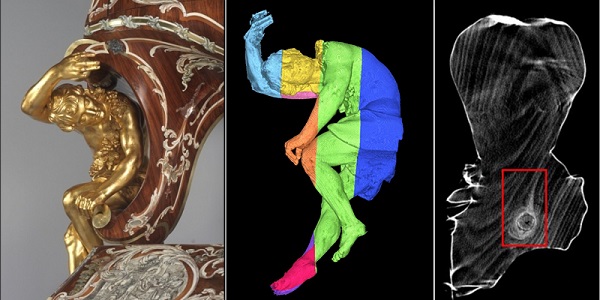
In the figures are shown respectively, a CT scan of a large wooden artworks (one of the lateral statues of "Doppio corpo" by Pietro Piffetti, Quirinale Palace, Rome)
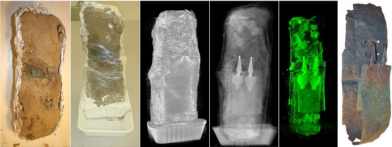
and a CT scan of a earth block from an archaeological excavation in Villalfonsina (CH, Italy) in collaboration with CCR "La Venaria Reale".